
Diagnostic Ultrasound
Introduction to diagnostic ultrasound
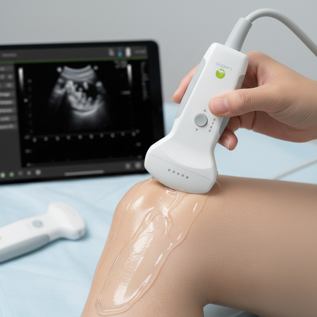
Diagnostic ultrasound—often called sonography—is a safe, versatile, and noninvasive imaging method that uses high-frequency sound waves to visualize internal structures in real time. Its safety profile and adaptability should inspire confidence in clinicians and students alike.
Ultrasound imaging has become one of the most versatile diagnostic modalities across modern medicine, with significant clinical applications. Its ability to deliver real-time images makes it indispensable for evaluating muscles, organs, blood flow, and soft tissues, and for guiding procedures with precision.
Key Takeaways
- No radiation exposure: Ultrasound relies entirely on sound waves, offering a safer alternative to modalities that use ionizing radiation.
- Real-time imaging: Clinicians can evaluate moving structures such as the heart, blood flow, tendons, and fetal motion instantly.
- Extremely versatile: Used across obstetrics, cardiology, vascular medicine, musculoskeletal imaging, abdominal diagnosis, thyroid evaluation, and procedural guidance.
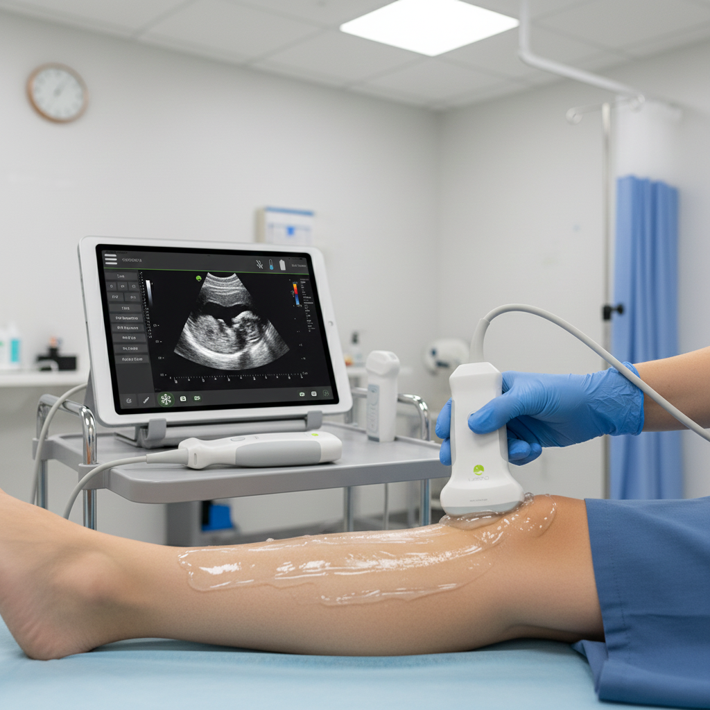
- Portable and accessible: Modern handheld and portable ultrasound units make rapid bedside imaging possible without transporting patients.
- Cost-effective: Ultrasound is significantly more affordable than CT or MRI while providing highly valuable diagnostic information.
- Precision in procedures: Ultrasound-guided biopsies, injections, aspirations, and catheter placements are safer and more accurate.
Clinical Applications Across Specialties
Obstetrics
- Fetal development monitoring: Ultrasound provides detailed images of fetal anatomy, heartbeat, growth curves, and movement.
- Placental health evaluation: Helps detect placental insufficiency, previa, and cord abnormalities.
- Safe for pregnancy: Zero radiation, making it the standard imaging tool for prenatal care.
Cardiology
- Assessing heart function: Echocardiography visualizes chambers, valves, contraction strength, and structural defects.
- Blood flow patterns: Doppler imaging reveals flow direction, velocity, and obstruction.
- Early detection: Useful for diagnosing valvular disease, heart failure, congenital defects, and pericardial effusions.
Musculoskeletal Imaging
- Soft tissue visualization: Ultrasound evaluates muscles, tendons, and ligaments for tears, inflammation, and degeneration.
- Dynamic movement testing: Real-time visualization allows clinicians to assess joints and soft tissue while the patient moves.
Abdominal Imaging
- Liver, kidney, gallbladder evaluation: Useful for diagnosing gallstones, liver disease, kidney stones, and organ enlargement.
- Non-invasive and fast: First-line choice for abdominal pain due to safety and speed.
Vascular Diagnostics
- Detecting blockages and DVT: Doppler ultrasound assesses venous and arterial flow, identifying clots or obstructions.
- PAD assessment: Measures blood flow in peripheral arteries to diagnose peripheral arterial disease.
Thyroid Imaging
- Characterizing thyroid nodules: Ultrasound differentiates solid vs. cystic masses and detects suspicious features.
- Biopsy guidance: Ensures accurate fine-needle aspiration for cytology.
Breast Ultrasound
- Evaluating lesions: Helps distinguish benign vs. malignant features, often used alongside mammography.
- Procedure support: Guides biopsies for precise tissue sampling.
Procedure Guidance
- Injections & aspirations: Real-time visualization increases safety in joint injections, nerve blocks, and cyst drainage.
- Biopsies: Ensures needle accuracy and reduces complication risks.
What to Look for in a High-Quality Ultrasound System
- High-resolution imaging: Clear visualization of tissue layers, organ borders and small structures improves diagnostic accuracy.
- Portability: Portable and handheld devices allow bedside imaging, essential for emergency rooms, mobile units, and remote care.
- User-friendly interface: Adjustable settings, intuitive menus, and touchscreen options enhance workflow.
- Advanced imaging modes: Color Doppler, spectral Doppler, 3D/4D imaging, and elastography provide added diagnostic value.
- AI-powered tools: Automated measurements, image optimization, and interpretation assistance streamline clinical workflow.
- Long battery life: Important for mobile providers, urgent care units, or field-based clinicians.
- DICOM compatibility: Ensures seamless image storage, retrieval, and integration with hospital PACS systems.
Science Behind Diagnostic Ultrasound
- Sound wave emission and return: The transducer sends high-frequency waves into the body; returning echoes form gray-scale images.
- Tissue reflection patterns:
- Fluids = dark anechoic areas
- Muscles = striated textures
- Organs = medium-gray patterns
- Bone and air = bright reflections
This allows clear differentiation between structural types.
- Speed of sound in tissues: Variations in reflection, refraction, and absorption contribute to image interpretation.
- Real-time imaging advantage: Superior for monitoring moving structures, guiding procedures, and immediate clinical decision-making.
Advanced Doppler Technology
- Measures blood flow: Doppler ultrasound evaluates direction, speed, and quality of circulation.
- Detects vascular disease: Helps identify narrowing, clots, turbulence, and vascular insufficiency.
- Color-coded mapping: Improves visualization for cardiology, obstetrics, and vascular surgery.
Example:
In fetal medicine, Doppler imaging analyzes umbilical cord flow to detect placental insufficiency or fetal distress.
Real-World Clinical Example
A cardiologist uses high-resolution Doppler ultrasound to analyze valve movement and blood flow direction in a patient with a suspected heart murmur. The real-time images help confirm the presence of regurgitation and guide immediate treatment decisions.
Historical Development of Diagnostic Ultrasound
- 1950s origin: Early ultrasound machines were large and basic, used mainly for detecting tissue boundaries.
- Advancements in resolution: Introduction of higher-frequency probes dramatically improved image clarity and diagnostic accuracy.
- Doppler revolution: Allowed detailed vascular assessments and cardiac imaging.
- Portable ultrasound evolution: Handheld devices brought imaging to ambulances, rural clinics, mobile teams, and bedside care.
- 3D/4D and elastography: Modern systems add depth, motion visualization, and tissue stiffness analysis for oncology, obstetrics, and liver disease.
Modern Innovations in Ultrasound Equipment
- AI-enhanced scanning: Offers automated image optimization, embryo measurement, and plaque characterization.
- Wireless probes: Reduce clutter and improve portability.
- Advanced cardiology features: Tissue Doppler, strain imaging, and speckle-tracking for assessing cardiac function.
- Dual-probe functionality: Allows rapid switching between linear and convex modes.
Latest Research
A recent Journal of Ultrasound in Medicine study validated the clinical impact of portable ultrasound systems in underserved regions, proving that modern handheld units can achieve diagnostic accuracy comparable to full-size hospital machines.
Mechanisms of Action
- Ultrasound wave penetration: Sound waves travel through tissue layers and reflect based on tissue density differences.
- Echo processing: The machine converts echo patterns into real-time images with varying brightness levels.
- Doppler frequency shifts: Used to analyze blood flow speed and direction by tracking moving red blood cells.
Case Studies
Real-World Example: Doppler in Obstetrics
Doppler ultrasound evaluates blood flow in the umbilical cord, detecting fetal distress, placental insufficiency, or high-risk pregnancy complications. It is a critical tool for ensuring fetal well-being during prenatal monitoring.
Ultrasound-Guided AC…
Patient Profile Clinical Presentation The patient presented with chronic AC joint pain, reduced mobility, and difficulty performing overhead work…
Detecting Early…
Patient Profile Clinical Scenario Given Megan’s increased risk profile, particularly with a twin gestation and prior preeclampsia, a Doppler…
Doppler Assessment…
Patient Profile Clinical Scenario Laura’s pregnancy was categorized as high-risk due to multiple factors. Her care team ordered a…
Monitoring Umbilical…
Patient Profile Clinical Scenario Rachel attended a routine prenatal scan. Due to her family history of hypertension, her obstetrician…